Difference between revisions of "CNN for Enhanced Glaucoma Detection By Avijit Singh"
| Line 19: | Line 19: | ||
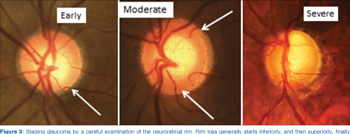
The Convolutional Neural Network used Fundus photographs to provide a 30-degree image of the optic nerve head, which is the structure in the posterior of the fundus that allows the exit of retinal cell axons and the entry and exit of blood vessels. The 30-degree image provides data for the network to quantify a number of glaucoma specific morphological features like neuroretinal rim loss. | The Convolutional Neural Network used Fundus photographs to provide a 30-degree image of the optic nerve head, which is the structure in the posterior of the fundus that allows the exit of retinal cell axons and the entry and exit of blood vessels. The 30-degree image provides data for the network to quantify a number of glaucoma specific morphological features like neuroretinal rim loss. | ||
| − | [[File:Glaucoma Progression.png]] | + | [[File: Glaucoma Progression.png]] |
But because the image may not take into account moving blood, fundus videos provide the network to analyze the videos in search of markers associated with blood flow. The Computer Aided Diagnosis systems played a role in making sure the CNN was accurate, reliable, and a fast diagnosis of glaucoma. The CNNs can facilitate autonomous classification based on features from thousands of fundus images. The specificity and sensitivity of these models range between 85 to 95% | But because the image may not take into account moving blood, fundus videos provide the network to analyze the videos in search of markers associated with blood flow. The Computer Aided Diagnosis systems played a role in making sure the CNN was accurate, reliable, and a fast diagnosis of glaucoma. The CNNs can facilitate autonomous classification based on features from thousands of fundus images. The specificity and sensitivity of these models range between 85 to 95% | ||
Revision as of 23:35, 21 October 2022
By Avijit Singh
A combined convolutional neural network and a recurrent neural network was developed that not only extracts the spatial features in a fundus image but also the temporal features embedded in a fundus video. A fundus camera or a retinal camera is a specialized low power microscope with an attached camera that is designed to photograph the interior surface of the eye: the retina, retinal vascularity, optic disk, macula, and the posterior pole. The fundus is the combination of the retina and the posterior pole all together. Fundus imaging is the process where the reflected light reflected is used to form a two-dimensional representation of the three-dimensional retina or the fundus (By changing dimensionality, this is similar to what we did in the Neural Network MatLab project).
What is Glaucoma?
Glaucoma is a leading cause of blindness worldwide. It is an eye disease that results in damage to the optic nerve and vision loss because of the buildup of intraocular pressure (IOP). The most common type of glaucoma is open-angle glaucoma where the drainage angle for fluid in the eye remains open. Open-angle glaucoma develops slowly over time and there is no pain associated with it, but peripheral vision may begin to decrease, followed by central vision and eventually blindness if left untreated. Closed-angle glaucoma is another less common type of glaucoma. Closed-angle glaucoma can present itself gradually or suddenly. If sudden, there may be severe eye pain, blurred vision, redness of the eye, and nausea. Vision loss due to glaucoma is permanent so the need to detect glaucoma early is of utmost importance.
Risk and Treatments
Glaucoma’s risk increases with age, high pressure in the eye, and with a family history of glaucoma. A normal eye pressure falls between 10 and 21 mm Hg with the average eye pressure being 15 mm Hg. Someone with glaucoma may have an eye pressure of above 21 mm Hg. If glaucoma is detected and treated early, it is possible to slow the progression of the disease with medication, laser treatment, or even surgery. In order to treat glaucoma, the pressure of the eye has to decrease.
There are about 80 million people worldwide with glaucoma and about 3 million Americans are affected by glaucoma. Glaucoma is the second leading cause of blindness behind cataracts. With a Convolutional Neural Network that is able to detect glaucoma early, many people could have their vision saved since vision loss caused by glaucoma is permanent.
The Convolutional Neural Network
The Convolutional Neural Network used Fundus photographs to provide a 30-degree image of the optic nerve head, which is the structure in the posterior of the fundus that allows the exit of retinal cell axons and the entry and exit of blood vessels. The 30-degree image provides data for the network to quantify a number of glaucoma specific morphological features like neuroretinal rim loss.
But because the image may not take into account moving blood, fundus videos provide the network to analyze the videos in search of markers associated with blood flow. The Computer Aided Diagnosis systems played a role in making sure the CNN was accurate, reliable, and a fast diagnosis of glaucoma. The CNNs can facilitate autonomous classification based on features from thousands of fundus images. The specificity and sensitivity of these models range between 85 to 95%
